welcome to anlianfitness!
Muscular System
The human body consists of over 600 muscles, which together account for about 40-50 percent of a person’s body weight. This article focuses on the skeletal muscles, providing you with a basic introduction to anatomical terminology and the names and functions of the relevant muscles throughout the entire body that are affected by resistance training.
MUSCULOSKELETAL SYSTEM
7/9/202410 min read
Sources
[1] Bryant, C. X., & Green, D. J. (2017). Ace Essentials of Exercise Science for Fitness Professionals. American Council on Exercise.
[2] Cleveland Clinic medical. (n.d.). How many muscles are in the human body?. Cleveland Clinic. https://my.clevelandclinic.org/health/body/muscles
[3] Crompton, R. Huw , Cummings, . Shane W. , Wood, . Bernard and Tangen, . Christopher (2024, January 21). human muscle system. Encyclopedia Britannica. https://www.britannica.com/science/human-muscle-system
[4] Seladi-Schulman, J. (2020, February 4). How many muscles are in the human body? plus a diagram. Healthline. https://www.healthline.com/health/how-many-muscles-are-in-the-human-body
[5] Taylor, T. (2021b, October 10). Interactive guide to the muscular system. Innerbody. https://www.innerbody.com/image/musfov.html
The human body consists of over 600 muscles, which together account for about 40-50 percent of a person’s body weight. The muscles are organized by tissue type. There are three types of muscle tissue: visceral, cardiac, and skeletal. Visceral and cardiac muscle act involuntarily, which means that their movements happen automatically to keep the body functioning properly. Visceral (or smooth) muscle is found inside of organ systems (e.g., stomach, intestines, and blood vessels), and cardiac muscle is only found in the heart, which is responsible for pumping blood throughout the body. Skeletal muscle is the only type of muscle tissue that acts voluntarily, which means that it is controlled consciously. Every physical action that a person consciously performs (e.g. speaking, walking, or lifting weights) requires skeletal muscle. When a muscle contracts, it allows movement of a specific area of the body.
Skeletal muscles are part of the musculoskeletal system because they work with the bones, tendons, and ligaments to support the body and its movements. Ligaments are fibrous connective tissues that attach bone to bone. The tendons attach the skeletal muscles to the bones throughout the body. Tendons are tough bands of dense regular connective tissue whose strong collagen fibers firmly attach muscles to bones. Tendons are under extreme stress when muscles pull on them, so they are very strong and are woven into the coverings of both muscles and bones. The functions of the skeletal muscles include enabling movement of the body, providing structural support, maintaining posture, generating heat to maintain body temperature, acting as a source of nutrients (e.g., amino acids), and serving as an energy source during starvation.
Before learning about the different muscles of the muscular system, it is important to be familiar with basic anatomical terminology. Below is an overview of anatomical, directional, positional, and regional terms as well as of the fundamental movements that are important to understand when learning about each muscle.
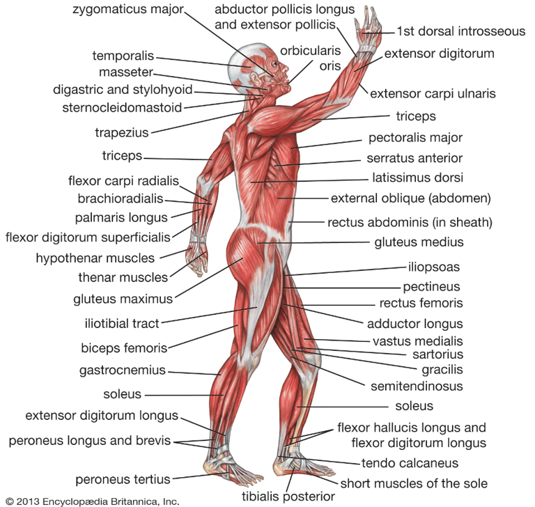
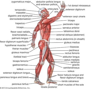
Figure 1. Diagram of human muscular system with muscles labeled [3].
Terminology
Anatomical, Directional, Positional, and Regional Terms
Anterior (ventral). Toward the front.
Posterior (dorsal). Toward the back.
Lateral. Away from the midline of the body.
Medial. Toward the midline of the body.
Inferior. Away from the head.
Superior. Toward the head.
Proximal. Toward the attached end of the limb, origin of the structure, or midline of the body.
Distal. Away from the attached end of the limb, origin of the structure, or midline of the body.
Superficial. External; located close to or on the body surface.
Deep. Internal; located further beneath the body surface.
Anatomical position. Standing with feet and palms facing towards the front.
Supine. Lying on your back, face up.
Prone. Lying on your stomach, face down.
Plantar. The sole or bottom of the feet.
Dorsal. The top surface of the feet and hands.
Palmar. The anterior or ventral surface of the hands.
Cervical. Regional term referring to the neck.
Thoracic. Regional term referring to the portion of the body between the neck and the abdomen; also known as the chest (thorax).
Lumbar. Regional term referring to the portion of the back between the abdomen and the pelvis.
Sagittal plane. A longitudinal (imaginary) line that divides the body or any of its parts into right and left sections.
Frontal plane. A longitudinal (imaginary) section that divides the body into anterior and posterior parts; lies at a right angle to th4e sagittal plane.
Transverse plane. Also known as the horizontal plane; an imaginary line that divides the body or any of its parts into superior and inferior sections.
Multiplanar. Describes a movement pattern that takes place in two or all three planes.
Terminology of Fundamental Movements (From Anatomical Position)
Sagittal Plane
Flexion. Decreasing the angle between two bones.
Extension. Increasing the angle between two bones.
Dorsiflexion. Moving the top of the foot toward the shin (only at the ankle joint).
Plantarflexion. Moving the sole of the foot downward; pointing the toes (only at the ankle).
Frontal Plane
Abduction. Motion away from the midline of the body (or part).
Adduction. Motion towards the midline of the body (or part).
Elevation. Moving to a superior position (only at the scapula).
Depression. Moving to an inferior position (only at the scapula).
Inversion. Lifting the medial border of the foot (only at the subtalar joint).
Eversion. Lifting the lateral border of the foot (only at the subtalar joint).
Transverse Plane
Rotation. Internal (inward) or external (outward) turning about the vertical axis of bone.
Pronation. Rotating the hand and wrist medially from the elbow; pronation of the foot is a combination of eversion and abduction, raising the lateral edge of the foot.
Supination. Rotating the hand and wrist laterally from the elbow; supination of the foot is a combination of inversion and adduction, raising the medial edge of the foot.
Horizontal flexion (adduction). From a 90-degree abducted shoulder or hip position, the humerus or femur, respectively, is flexed (adducted) in toward the midline of the body in the transverse plane.
Horizontal extension (abduction). The return of the humerus or femur from horizontal flexion (adduction).
Multiplanar
Circumduction. Motion that describes a cone; combines flexion, extension, abduction, and adduction in sequence.
Opposition. Thumb movement unique to humans and primates.
Muscular Anatomy
Skeletal muscles are named according to their location; origin and insertion; number of origins; shape, size, and direction; and function.
Location. Some skeletal muscles are named based on their anatomical region. For example, the rectus abdominis and transverse abdominis muscles are found in the abdominal region. Some muscles are named after the part of the bone that they are attached to (e.g., tibialis anterior). Other muscles use a hybrid of these two, like the brachioradialis, which is named after a region (brachial) and a bone (radius).
Origin and insertion. Some skeletal muscles are named according to their connection to a stationary bone (origin) and a moving bone (insertion). These muscles become very easy to identify once you know the names of the bones that they are attached to. Examples of this type of muscle include the sternocleidomastoid (connecting the sternum and clavicle to the mastoid process of the skull) and the occipitofrontalis (connecting the occipital bone to the frontal bone).
Number of origins. Some skeletal muscles connect to more than one bone or to more than one place on a bone, and therefore have more than one origin. A muscle with two origins is called a biceps muscle. A muscle with three origins is called a triceps muscle. Finally, a muscle with four origins is called a quadriceps muscle.
Shape, size, and direction. Skeletal muscles are also classified by their shapes. For example, the deltoids have a delta or triangular shape, and the rhomboid major is a rhombus or diamond shape. The size of the muscle can be used to distinguish between two muscles found in the same region. The gluteal region contains three muscles differentiated by size, i.e., the gluteus maximus (large), gluteus medius (medium), and gluteus minimus (smallest). Finally, the direction in which the muscle fibers run can be used to identify a muscle. In the abdominal region, there are several sets of wide, flat muscles. The muscles whose fibers run straight up and down are the rectus abdominis, the ones running transversely (left to right) are the transverse abdominis, and the ones running at an angle are the obliques.
Function. Skeletal muscles are sometimes classified by the type of function that they perform. Most of the muscles of the forearms are named based on their function because they are located in the same region and have similar shapes and sizes. For example, the flexor group of the forearm flexes the wrist and the fingers. The supinator is a muscle that supinates the wrist by rolling it over to face palm up. In the leg, there are muscles called adductors whose role is to adduct (pull together) the legs.
The following diagrams illustrate all the relevant muscles in the human body that you should be aware of, because when you lift weights or train with resistance, these muscles are most likely being affected. The following diagrams are divided into the muscles of the upper extremity, muscles of the trunk, and muscles of the lower extremity. Moreover, the primary functions of the major muscles that act at each region and body part are listed.
Primary Functions of the Major Muscles That Act at the Shoulder Girdle
Trapezius. (Upper): upward rotation and elevation of scapula; (middle): upward rotation and adduction of scapula; and (lower): depression of scapula.
Levator scapulae. Elevation of scapula.
Rhomboid major and minor. Adduction, downward rotation, and elevation of scapula.
Pectoralis minor. Stabilization, depression, downward rotation, and abduction of scapula.
Serratus anterior. Stabilization, abduction, and upward rotation of scapula.
Muscles of the Upper Extremity
Primary Functions of the Major Muscles That Act at the Shoulder Joint
Pectoralis major. Flexion, extension, adduction, internal rotation, and horizontal adduction.
Deltoid. (Entire muscle): abduction; (anterior fibers): flexion, internal rotation, and horizontal adduction; (posterior fibers): external rotation and horizontal abduction.
Latissimus dorsi. Extension, adduction, horizontal abduction, and internal rotation.
Rotator cuff. (Infraspinatus and teres minor): external rotation; (subscapularis): internal rotation; (supraspinatus): abduction. All contribute to the stability of the humeral head.
Teres major. Extension, adduction, and internal rotation.
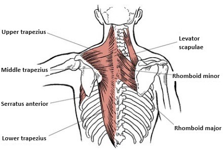
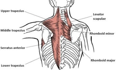

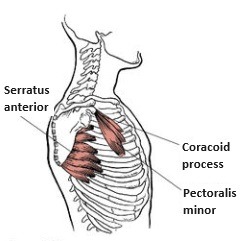
Figure 2. Superficial and deep muscles that act at the scapulothoracic articulation [1].
Figure 3. Anterior shoulder girdle muscles [1].

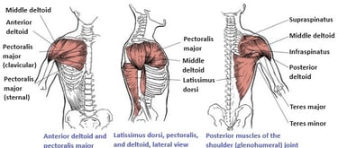
Figure 4. Superficial shoulder muscles [1].
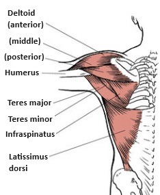

Figure 6. Superficial musculature of the superior and inferior shoulder joint [1].
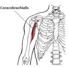

Figure 7. Deep muscle that originates on the shoulder blade and assists in flexion and adduction of the arm [1].
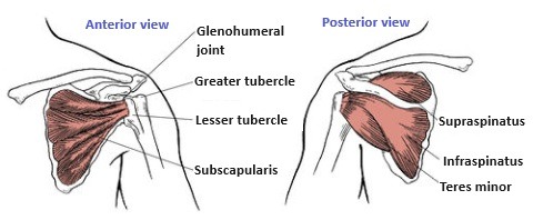
Figure 5. Rotator cuff muscles [1].
Primary Functions of the Major Muscles That Act at the Elbow and Forearm
Biceps brachii. Flexion at elbow and supination at forearm.
Brachialis. Flexion at elbow.
Brachioradialis. Flexion at elbow and supination at forearm.
Triceps brachii. Extension at elbow and arm extension (long head).
Pronator teres. Flexion at elbow and pronation at forearm.
Pronator quadratus. Pronation at forearm.
Supinator. Supination at forearm.
Primary Functions of the Major Muscles That Act at the Wrist
Flexor carpi radialis. Flexion.
Flexor carpi ulnaris. Flexion.
Extensor carpi radialis longus. Extension.
Extensor carpi ulnaris. Extension.
Palmaris longus. Flexion.

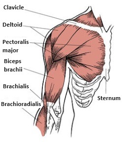
Figure 8. Superficial musculature of the anterior chest, shoulder, and arm [1].
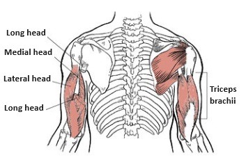
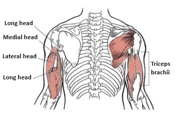
Figure 9. Triceps: long, medial, and lateral heads [1].
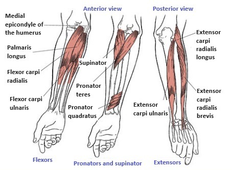
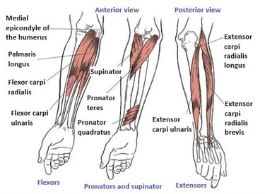
Figure 10. Muscles of the wrist [1].
Muscles of the Trunk
Primary Functions of the Major Muscles That Act at the Trunk
Rectus abdominis. Flexion and lateral flexion of the trunk.
External oblique. Contralateral rotation, lateral flexion, and forward flexion (both sides).
Internal oblique. Ipsilateral rotation, lateral flexion, and forward flexion (both sides).
Transverse abdominis. Compresses abdomen.
Erector spinae. Extension (both sides) and lateral flexion.
Primary Functions of the Major Muscles That Act at the Hip
Iliopsoas: iliacus and psoas major and minor. Flexion and external rotation.
Rectus femoris. Flexion.
Gluteus maximus. Extension and external rotation. (Superior fibers): abduction.
Biceps femoris. Extension, abduction, and slight external rotation.
Semitendinosus. Extension, adduction, and slight internal rotation.
Semimembranosus. Extension, adduction, and slight internal rotation.
Gluteus medius and minimus. (All fibers): abduction; (anterior fibers): internal rotation; (posterior fibers): external rotation.
Adductor magnus. Adduction.
Adductor brevis and longus. Adduction.
Tensor fasciae latae. Flexion, abduction, and internal rotation.
Sartorius. Flexion and external rotation of the hip, flexion of the knee.
Pectineus. Flexion, adduction, and external rotation.
Gracilis. Adduction.
Piriformis. External (lateral) rotation.
Obturator externus and internus. External (lateral) rotation.
Gemellus superior and inferior. External (lateral) rotation.
Quadratus femoris. External (lateral) rotation.
Primary Functions of the Major Muscles That Act at the Knee
Rectus femoris. Extension (most effective when the hip is extended).
Vastus lateralis, intermedius, and medialis. Extension.
Biceps femoris. Flexion and external rotation.
Semitendinosus. Flexion and internal rotation.
Gracilis. Flexion.
Sartorius. Flexion and external rotation of the hip, flexion of the knee.
Popliteus. Knee flexion, internal rotation of the lower leg to unlock the knee.
Primary Functions of the Major Muscles That Act at the Ankle and Foot
Anterior tibialis. Dorsiflexion at ankle and inversion at foot.
Peroneus longus and brevis. Plantarflexion at ankle and eversion at foot.
Gastrocnemius. Plantarflexion at ankle and flexion at knee.
Soleus. Plantarflexion at ankle.
Posterior tibialis. Plantarflexion at ankle and inversion at foot.
Extensor hallucis longus. Dorsiflexion and inversion of foot, extension of great toe.
Extensor digitorum longus. Dorsiflexion and eversion of foot, extension of toes 2 through 5.
Peroneus tertius. Dorsiflexion and eversion of foot.
Plantaris. Flexion of knee and plantarflexion of foot.
Flexor hallucis longus. Flexion of great toe, plantarflexion and inversion of foot.
Flexor digitorum longus. Flexion of toes 2 through 5, plantarflexion and inversion of foot.
Muscles of the Lower Extremity
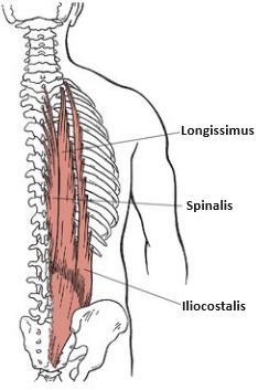

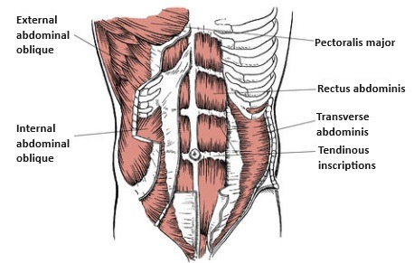
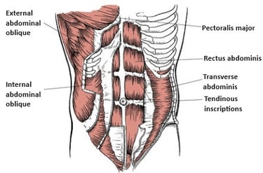
Figure 11. Muscles of the posterior chain (erector spinae) [1].
Figure 12. Muscles of the abdominal wall [1].
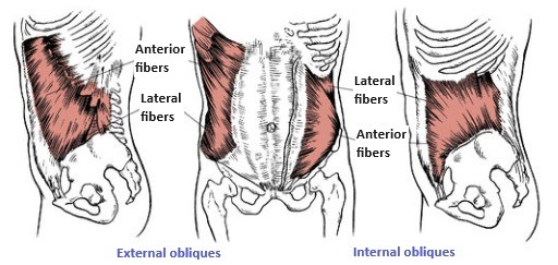
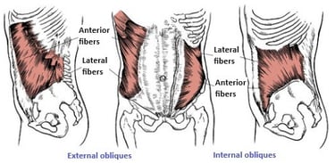
Figure 13. External and internal obliques [1].
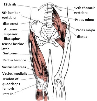

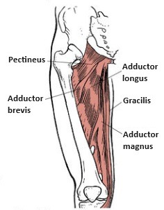

Figure 14. Anterior musculature of the hip and knee [1].
Figure 15. Medial muscles of the hip that are responsible for adduction [1].

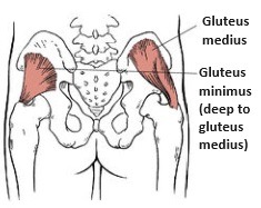

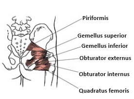
Figure 16. Abductors of the posterior hip [1].
Figure 17. Deep rotators of the hip [1].

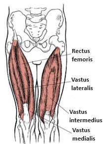
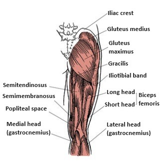

Figure 18. Quadriceps muscles [1].
Figure 19. Posterior musculature of the hip and knee [1].

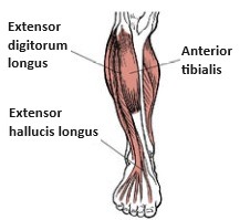

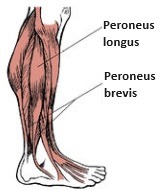

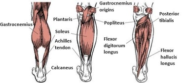
Figure 20. Anterior tibial compartment muscles [1].
Figure 21. Lateral tibial compartment muscles [1].
Figure 22. Posterior tibial compartment muscles [1].
Copyright © 2024-2025 AnlianFitness. All rights reserved.
Follow us on social media.